Upon pathogen invasion, myeloid cells are rapidly recruited to local tissues via various chemokine receptors where they are activated for phagocytosis and secrete inflammatory cytokines, thus playing a major role in innate immunity. Thus, CD Genomics provides myeloid cell protein profiling by CyTOF to facilitate further insight into the complex myeloid cell composition in the lesion in question.
Granulocytes and monocytes, collectively known as bone marrow cells, are descended from a common progenitor of hematopoietic stem cells differentiated in the bone marrow. Monocytes eventually mature into macrophages in various tissues where they may exhibit unique, tissue-dependent morphology and specific functions. Monocytes may also differentiate into dendritic cells (DCs) in lymphoid organs and Langerhans cells in the skin, which act as specialized antigen-presenting cells. Granulocytes are the first cells to be recruited to local sites upon pathogen invasion and provide a direct line of defense against infection in tissues.
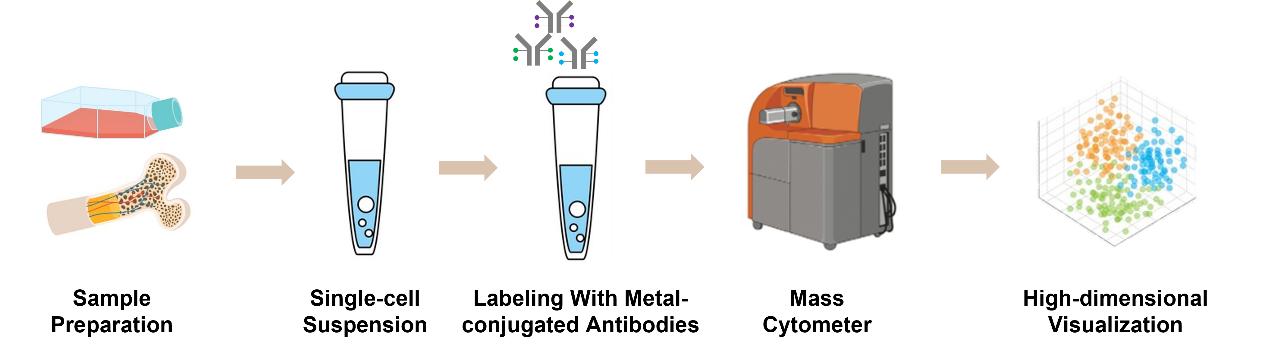
The development of mass spectrometry, a technique that combines flow cytometry and mass spectrometry, has enabled the detection of up to 40 protein reads in single cells. This has contributed to the understanding of the phenotypic diversity of bone marrow cells found in vitro and in vivo. These studies have identified the phenotypic characteristics of myeloid cells found in different types of tissues and at different stages of disease development with unprecedented resolution.
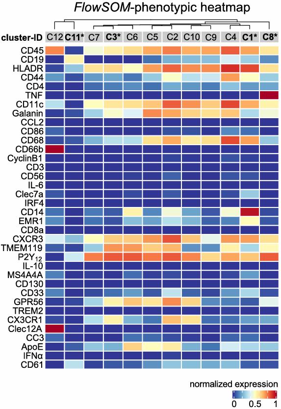 Fig.1 Heat map clusters show the expression levels of all 36 markers used for cluster analysis. (Böttcher, C., et al., 2020)
The application of technology at the single-cell level provides researchers with a new perspective on the complexity of multiple diseases. CyTOF utilizes the principle of mass spectrometry to simultaneously detect multiple indicators at the single-cell level. CyTOF allows for high-throughput, multi-parameter detection of large numbers of cells, which in turn allows for accurate immunophenotyping of cell populations. Therefore, CD Genomics offers CyTOF-based services for the protein profiles diversity analysis of myeloid cells.
CD Genomics has specialized in the single-cell analysis of immune cells for many years and has gained extensive experience in CyTOF and immunomics. We ensure that your project is carried out in the best possible way and that reliable data are obtained. Please contact us for more information.


